Giant dermoid cyst mimicking posterior fossa tumors in a child: A Case Report and Review of the Literature
DOI:
https://doi.org/10.46900/apn.v2i2(May-August).45Keywords:
intracranial dermoid cyst, posterior fossa, dermal sinus, magnetic resonance imagingAbstract
Introduction: Intracranial dermoid cysts are rare, congenital and, benign lesions. The etiology of these lesions is related to an embryonic defect during neurulation.
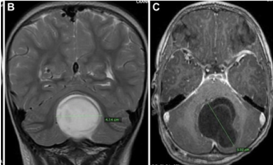
Case presentation: The present study describes a case of a 3-year-old girl with a giant cerebellar dermoid cyst, which initially manifested as hydrocephalus.
Discussion: We discuss its epidemiological characteristics as well as diagnostic and therapeutic management. The combination of high clinical suspicion, anamnesis, thorough physical examination, and adequate interpretation of neuroimaging data is crucial for the early diagnosis and timely therapeutic intervention for such cysts.
Conclusion: Surgical approach involving complete lesion resection considerably improves prognosis.
Downloads

Downloads
Published
How to Cite
Issue
Section
License
Copyright (c) 2020 Leopoldo Mandic Ferreira Furtado, José Aloysio da Costa Val Filho, Bruno Lacerda Sandes, Plínio Duarte Mendes, Patrícia Salomé Gouvea Braga

This work is licensed under a Creative Commons Attribution 4.0 International License.

When publishing in Archives of Pediatric Neurosurgery journal, authors retain the copyright of their article and agree to license their work using a Creative Commons Attribution 4.0 International Public License (CC BY 4.0), thereby accepting the terms and conditions of this license (https://creativecommons.org/licenses/by/4.0/legalcode).
The CC BY 4.0 license terms applies to both readers and the publisher and allows them to: share (copy and redistribute in any medium or format) and adapt (remix, transform, and build upon) the article for any purpose, even commercially, provided that appropriate credit is given to the authors and the journal in which the article was published.
Authors grant Archives of Pediatric Neurosurgery the right to first publish the article and identify itself as the original publisher. Under the terms of the CC BY 4.0 license, authors allow the journal to distribute the article in third party databases, as long as its original authors and citation details are identified.


