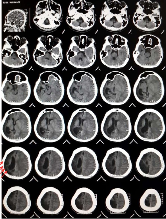
From Sinus Infection to Subdural Empyema: A Pediatric Epileptic Case with Imaging Challenges and Misdiagnosis
DOI:
https://doi.org/10.46900/apn.v8i3.384Keywords:
Subdural Empyema, Epilepsy, Pansinusitis, Traumatic Brain InjuryAbstract
Background: The unique anatomical and physiological features of the pediatric spine predispose children to a distinct form of spinal cord trauma known as SCIWORA (Spinal Cord Injury Without Radiographic Abnormality). This condition occurs predominantly in children and is characterized by clinical evidence of spinal cord injury in the absence of detectable abnormalities on conventional imaging modalities, including plain radiography and MRI. Recent studies indicate that multilevel spinal cord involvement in pediatric SCIWORA cases may be more frequent than previously recognized, and some patients may ultimately require surgical management.
Methods: A retrospective analysis was performed on 80 children aged 4–18 years who were treated for SCIWORA at the Pediatric Neurosurgery Department in Omsk between 2008 and 2022. The mechanisms of injury were evaluated, and neurological status was assessed using the Frankel scale to determine the severity of neurological deficits. Injury severity was additionally classified according to the criteria proposed by Zwimpfer T.J. (1990). All patients underwent spinal radiography in two standard projections as well as MRI examination. In twenty-eight cases, electromyography (EMG) studies were performed.
Results: In two patients, clinical manifestations of L4–L5 radiculopathy were observed despite the absence of lumbar spinal trauma. All patients demonstrated favorable outcomes following conservative treatment.
Conclusions: Pediatric SCIWORA remains a relatively rare yet potentially severe form of spinal cord injury. In children with SCIWORA syndrome, multifocal spinal cord involvement is possible, and the degree of neurological recovery largely depends on the presence and severity of MRI-detected abnormalities.
Downloads
Downloads
Published
How to Cite
Issue
Section
Categories
License
Copyright (c) 2026 Yuriz Bakhtiar, Rafi Ilmansyah; Muhammad Sinatraya Caropeboka; Ifandias Gian Abhista, Afrizal Hasan, Yuliana Masnita Dongoran

This work is licensed under a Creative Commons Attribution 4.0 International License.

When publishing in Archives of Pediatric Neurosurgery journal, authors retain the copyright of their article and agree to license their work using a Creative Commons Attribution 4.0 International Public License (CC BY 4.0), thereby accepting the terms and conditions of this license (https://creativecommons.org/licenses/by/4.0/legalcode).
The CC BY 4.0 license terms applies to both readers and the publisher and allows them to: share (copy and redistribute in any medium or format) and adapt (remix, transform, and build upon) the article for any purpose, even commercially, provided that appropriate credit is given to the authors and the journal in which the article was published.
Authors grant Archives of Pediatric Neurosurgery the right to first publish the article and identify itself as the original publisher. Under the terms of the CC BY 4.0 license, authors allow the journal to distribute the article in third party databases, as long as its original authors and citation details are identified.