How unusually located can a distal tip of a VP shunt be?
DOI:
https://doi.org/10.46900/apn.v5i1.105Keywords:
Hepatic pseudocyst, Intrahepatic pseudocyst, VP shunt, Ventriculoperitoneal shuntAbstract
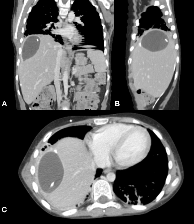
The clinical image is a computed tomography (CT) scan of the abdomen in the coronal (A), sagittal (B) and axial (C) incidences. We observe an intrahepatic (segment VII) CSF containing cyst along with the VP shunt distal tip. A 12-year-old male patient underwent a VP shunt due to hydrocephalus from tumoral etiology (craniopharyngioma). The patient had already had the shunt for 7 months when he started with severe abdominal pain on the right flank. An abdominal CT scan disclosed this rare finding. He underwent surgery to revise the shunt with repositioned towards inferior left quadrant of the abdomen. CSF analysis showed that there were no signs of infection. The patient did well without further complications. We reviewed the medical literature worldwide and found only 13 reports of intrahepatic CSF pseudocysts [1-5].
Received: 26 June 2021
Accepted: 12 July 2021
Published: 05 August 2021
Downloads
References
1. Arsanious D, Sribnick E. Intrahepatic Cerebrospinal Fluid Pseudocyst: A Case Report and Systematic Review. World Neurosurg. 2019 May;125:111-116. doi: 10.1016/j.wneu.2019.01.150. Epub 2019 Feb 2. PMID: 30721775.
2. Dabdoub CB, Fontoura EA, Santos EA, Romero PC, Diniz CA. Hepatic cerebrospinal fluid pseudocyst: A rare complication of ventriculoperitoneal shunt. Surg Neurol Int. 2013 Dec 27;4:162. doi: 10.4103/2152-7806.123783. PMID: 24523999; PMCID: PMC3908696.
3. Banka S, Johnson K, Sgouros S. Liver cyst caused by the peritoneal catheter of a cerebrospinal fluid shunt. Pediatr Neurosurg. 2007;43(4):343-4. doi: 10.1159/000103320. PMID: 17627156.
4. Kumar MM, Jeyabalachandran M, Sekar S. Intrahepatic cyst-a complication of ventriculoperitoneal shunt. J Indian Med Assoc 1995;93:403.
5. Rana SR, Quivers ES, Haddy TB. Hepatic cyst associated with ventriculo-peritoneal shunt in a child with bra tumor. Child’s Nerv Syst 1985;1:349–51.

Additional Files
Published
How to Cite
Issue
Section
License
Copyright (c) 2021 Marcos Rodrigo Pereira Eismann, Daniel Santos Sousa, Letícia Novak Crestani , Charles Kondageski

This work is licensed under a Creative Commons Attribution 4.0 International License.

When publishing in Archives of Pediatric Neurosurgery journal, authors retain the copyright of their article and agree to license their work using a Creative Commons Attribution 4.0 International Public License (CC BY 4.0), thereby accepting the terms and conditions of this license (https://creativecommons.org/licenses/by/4.0/legalcode).
The CC BY 4.0 license terms applies to both readers and the publisher and allows them to: share (copy and redistribute in any medium or format) and adapt (remix, transform, and build upon) the article for any purpose, even commercially, provided that appropriate credit is given to the authors and the journal in which the article was published.
Authors grant Archives of Pediatric Neurosurgery the right to first publish the article and identify itself as the original publisher. Under the terms of the CC BY 4.0 license, authors allow the journal to distribute the article in third party databases, as long as its original authors and citation details are identified.


