Pre- and postoperative intracranial volumes in children with non-syndromic craniosynostosis using 3D volumetric technique
DOI:
https://doi.org/10.46900/apn.v6i3.263Keywords:
Intracranial pressure, Craniosynostosis, Craniosynostosis, Intracranial volume, fronto-orbital advancement surgery, Renier's h surgery.Abstract
Introduction: Corrective surgery for craniosynostosis presents several challenges to students during their training, with obtaining practical experience being one of the main obstacles. The neurocranium is formed by the bony structures that surround the brain, thus ensuring its protection. During the first years of life, the baby's skull grows exponentially. Understanding the normal growth and development of the cranial shape is essential for monitoring cranial development, detecting abnormalities, and evaluating the long-term results of craniosynostosis surgery.
Objectives: To study the cranial volume gained after surgical treatment of craniosynostosis using 3D printing technology.
Methodology: 26 patients who underwent craniosynostosis surgery, from 2019 to 2022 were selected. Preoperative, immediate, and late postoperative (3 months) tomography examination was performed. Reconstructing the exams using the Blender program with cranial volume calculation. 3D printing of skulls using Sethi 3D printer. Evaluation of results using Student's t-test with independent samples.
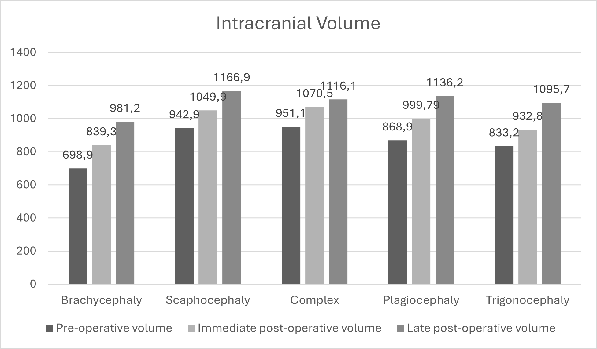
Results: The study involved 26 patients, with ten presenting scaphocephaly treated with Renier's "H" cranial remodeling, five with trigonocephaly, five with plagiocephaly, and two with brachycephaly treated with front-orbital advancement (FOA). Cranial volumes were measured before and after surgery. For patients undergoing Renier's "H" treatment, there was an average increase of 224 cm³ between the late postoperative and preoperative stages, with smaller differences between the immediate postoperative and preoperative stages. For those treated with FOA, the average difference between late postoperative and preoperative stages was 138.8 cm³, while between immediate postoperative and preoperative stages was 129.7 cm³.
Conclusion: Cranial volume gained by patients undergoing Renier's h technique and Fronto-orbital advancement is significant, showing that it may be possible to use less invasive techniques to take advantage of patients' natural volumetric gain.
Downloads

Downloads
Published
How to Cite
Issue
Section
Categories
License
Copyright (c) 2024 Gustavo Sampaio, Linoel Curado Valsechi, Renne Peres dos Santos Silva, Antonio Soares Souza

This work is licensed under a Creative Commons Attribution 4.0 International License.

When publishing in Archives of Pediatric Neurosurgery journal, authors retain the copyright of their article and agree to license their work using a Creative Commons Attribution 4.0 International Public License (CC BY 4.0), thereby accepting the terms and conditions of this license (https://creativecommons.org/licenses/by/4.0/legalcode).
The CC BY 4.0 license terms applies to both readers and the publisher and allows them to: share (copy and redistribute in any medium or format) and adapt (remix, transform, and build upon) the article for any purpose, even commercially, provided that appropriate credit is given to the authors and the journal in which the article was published.
Authors grant Archives of Pediatric Neurosurgery the right to first publish the article and identify itself as the original publisher. Under the terms of the CC BY 4.0 license, authors allow the journal to distribute the article in third party databases, as long as its original authors and citation details are identified.


