Differential diagnosis of bulging scalp: beyond cephalohematoma and subgaleal hematoma
DOI:
https://doi.org/10.46900/apn.v5i3.218Keywords:
delayed subaponeurotic fluid collection; magnetic resonance imaging; ultrassound; infant scalp collectionAbstract
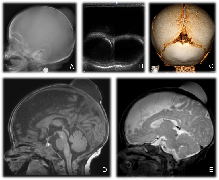
A 30-day-old infant was brought to the pediatric emergency room with a bulge in the scalp in the parieto-occipital region, mobile and without associated phlogistic signs. He had no history of fever or any other complaints. The mother reported that the bulge appeared at 20 days of life and showed progressive growth. There was no history of trauma. The boy was born at term by cesarean delivery due to cephalopelvic disproportion.
For initial evaluation, skull X-ray and cerebral ultrasound were performed (Figure 1). A magnetic resonance imaging and tomography of the brain were also performed (Figure 1).
Faced with the diagnostic challenge, neurosurgery proceeded with drainage of the collection and biopsy of the galea. No malignant cells were found and the biopsy only found an inflammatory change. Given these findings, the diagnosis of delayed subaponeurotic fluid collection (DSFD) was made. There was no recurrence of the collection and the patient had a good evolution.
DSFD are a rare condition, which occurs spontaneously between the 4th and 18th week of life, and makes the differential diagnosis with cephalohematoma, caput succedeneum and subgaleal hematoma [1]. Its pathophysiology is not fully elucidated; however, most reports share a similar history of prolonged or instrumented delivery [1,2]. Patients are otherwise healthy and with no history of trauma [2].
The management of DSFD is conservative and the natural history is spontaneous resolution within 1 to 2 months [1,2].
Downloads
References
Wang S, Drake J, and Kulkarni AV. Management and outcome of spontaneous subaponeurotic fluid collections in infants: the Hospital for Sick Children experience and review of the literature. Journal of Neurosurgery: Pediatrics 18.4 (2016): 442-447.
Faried A, Imron A, Aliyannissa A, Indrawati D. Delayed subaponeurotic fluid collection on an infant’s head: Underreported case and review of the literature. Surgical Neurology International 12 (2021).
Downloads
Additional Files
Published
How to Cite
Issue
Section
License
Copyright (c) 2023 Lillian Gonçalves Campos, Tassia Andrea Duraes Prioste, Jorge Wladimir Junqueira Bizzi

This work is licensed under a Creative Commons Attribution 4.0 International License.

When publishing in Archives of Pediatric Neurosurgery journal, authors retain the copyright of their article and agree to license their work using a Creative Commons Attribution 4.0 International Public License (CC BY 4.0), thereby accepting the terms and conditions of this license (https://creativecommons.org/licenses/by/4.0/legalcode).
The CC BY 4.0 license terms applies to both readers and the publisher and allows them to: share (copy and redistribute in any medium or format) and adapt (remix, transform, and build upon) the article for any purpose, even commercially, provided that appropriate credit is given to the authors and the journal in which the article was published.
Authors grant Archives of Pediatric Neurosurgery the right to first publish the article and identify itself as the original publisher. Under the terms of the CC BY 4.0 license, authors allow the journal to distribute the article in third party databases, as long as its original authors and citation details are identified.